The precise quantification of the proteins in a sample is essential for their study in an infinite number of research fields.
While the quantification of a specific protein can be carried out using assays such as Western Blot or ELISA, by mass spectrometry (MS) or by other methods such as those based on nanoparticles, there are assays that allow quantifying the total protein concentration present in a sample.
This last case is the one we focus on by compiling a brief description of the 5 main methods to quantify total proteins.
In Abyntek Biopharma we help you find the antibody, protein or ELISA kit you need.
Contact us or browse our catalogue with more than 7 million reagents for research.
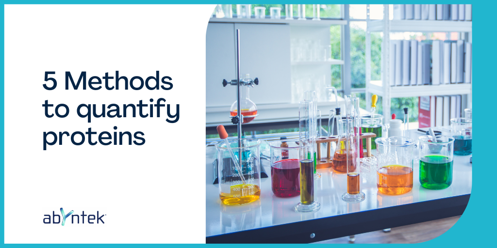
5 Methods to quantify proteins
1.- BRADFORD OR COOMASIE BLUE TEST
- Concentration range: 20-2000 ug/ml
- Characteristics:
- Method described in 1976.
- Negatively charged Coomasie blue stain binds positively charged proteins.
- Binds to arginine, tryptophan, tyrosine, histidine and phenylalanine residues.
- Staining in solution is red and absorbs at 465 nm, while when bound to the basic amino acids of a protein it turns blue and absorbs at 595 nm.
- The absorbance measurement is compared to the values of a standard curve to determine the protein concentration of the sample.
- Advantages:
- It is characterized by being a fast, simple and compatible method with the reducing agents used to stabilize proteins in solution.
- Limitations:
- Not valid for detecting <3kDa proteins.
- It is incompatible with detergents such as SDS or Triton X-100.
2.- Lowry test
- Concentration range: 10-1000 ug/ml.
- Characteristics:
- One of the most used methods to quantify proteins, was developed in 1951.
- The reaction occurs in two steps:
- Formation of Cu-N complexes (present in the protein).
- Tyrosine and tryptophan complexes react with Folin -Ciocalteau reagent, producing a blue-green colour that absorbs between 650 and 750 nm.
- The absorbance measurement is compared to the values of a standard curve to determine the protein concentration of the sample.
- Advantages:
- It is a very sensitive and accurate assay.
- Limitations:
- It is incompatible with certain commonly used chemical reagents such as Tris, EDTA, DDT, 2-mercaptoethanol, carbohydrates, etc.
3.- BICINCHONINIC ACID (BCA)
- Concentration range: 20-2000 ug/ml
- Characteristics:
- Developed in 1985.
- Colorimetric test where the absorbance will be proportional to the protein concentration present in the sample.
- As well as in the Lowry test, a reaction occurs in two steps:
- Formation of protein-copper ion complexes.
- Formation of a Cu-BCA chelate that gives rise to an intense purple coloration that absorbs at 562 nm.
- Advantages:
- It is recommended method in the case of samples containing >5% detergents or denaturing agents such as urea or guanidium chloride.
- Sensitivity comparable to that of the Lowry method.
- Limitations:
- Samples containing substances that interact with copper, such as ammonia, as well as EDTA, reducing sugars, or lipids, may interfere with this assay.
- Colour is not stable over time; time between analysis and absorbance reading needs to be carefully controlled.
4.- BIURET METHOD
- Concentration range: 0.373-80 g/L
- Characteristics:
- It is based on the formation of a coloured complex between the Cu2+ and the NH groups of the peptide bonds in a basic medium. 1Cu2+ complexes with 4 NH. The colour intensity is directly proportional to the amount of protein (peptide bonds) and the reaction is quite specific, so few substances interfere.
- The absorbance of the colour produced is read at 540 nm.
- The sensitivity of the method is very low and it is only recommended for the quantification of proteins in highly concentrated preparations (for example, in serum).
- The Biuret reagent is prepared as follows:
- Dissolve 3.8 g of CuSO4.5H2O and 6.7 g of NaEDTA in 700 ml of H2O.
- While stirring add 200 ml of 5N NaOH and then 1 g of KI as stabilizer.
- Store in a plastic bottle.
- Advantages:
- It is a fast method, it can be completed in less than 30 min.
- It is the simplest method to analyse proteins.
- Limitations:
- Insensitive method.
- Requires at least 2 to 4 mg of protein per test.
5.- ULTRAVIOLET (UV) ABSORPTION
- Concentration range: 0,1-100 ug/ml.
- Characteristics:
- By measuring the characteristic absorption of tryptophan and tyrosine at 280 nm, this method estimates the amount of protein present in the sample.
- Advantages:
- Fast and relatively sensitive method.
- Limitations:
- It is incompatible with protein extraction methods that use detergents or denaturing agents.
- It is not a specific method for proteins, since other compounds that could be in the sample also absorb at 280 nm (such as alcohols or nucleic acids, among others).
- A pure system is required to be able to use this method.
¿Do you know our search engine for antibodies, proteins or Elisa kits for research?
Our scientific team will advise you without obligation on the reagent that best suits your project. Contact us here

