In this Flow cytometry protocol we describe a powerful tool that has applications in multiple disciplines such as immunology, virology, molecular biology, cancer biology and infectious disease monitoring.
Flow cytometry is a technology that rapidly analyzes single cells or particles as they flow past single or multiple lasers while suspended in a buffered salt-based solution. Each particle is analyzed for visible light scatter and one or multiple fluorescence parameters.
It allows for the simultaneous characterization of mixed populations of cells from blood and bone marrow as well as solid tissues that can be dissociated into single cells such as lymph nodes, spleen, mucosal tissues, solid tumors etc. In addition to analysis of populations of cells, a major application flow cytometry is sorting cells for further analysis.
The instrumentation used for flow cytometry has evolved over the last several decades. Multiple laser systems are common as are instruments that are designed for specific purposes, such as systems with 96-well loaders designed for bead analysis, systems that combine microscopy and flow cytometry and systems that combine mass spectrometry and flow cytometry.
This Flow Cytometry protocol includes:
- Introduction
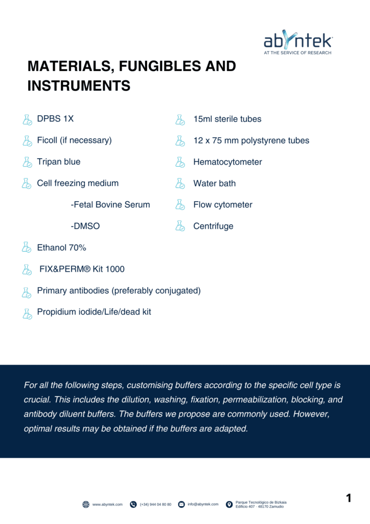
- Materials, Fungibles and Instruments
- Sample Preparation
- Thawing of cell suspensions
- Fixation, Permebilization and Staining
- Visibility Staning
- Controls
- Troubleshooting
Related content
The complete guide to Inmunohistochemistry
About Abyntek
At Abyntek Biopharma we have more than 15 years of experience giving the best possible service to researchers. That’s why we put all our know-how and experience both into helping researchers to find the perfect reagent for their research. And why we have put all the experience of our scientific team to create support documents such as these to guide and help researchers on their day to day.

